Insight 23 — When the Radiolucency Under a Restoration Is Not Caries
Clinical explanation
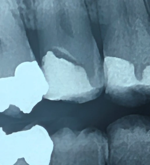
On bitewing radiographs, seeing a radiolucency adjacent to restorations can immediately lead the mind toward secondary caries.
However, in some cases a radiolucent area with defined borders is seen under the restoration whose origin is not caries, but is related to the restorative materials.
This situation is usually seen when:
flowable composite has been used as the underlying layer
glass ionomer or RMGI has been placed as a base
or a resin layer with low radiopacity has been placed on the floor of the cavity
Key radiographic point
When the radiolucency:
has smooth, regular borders
has a uniform thickness
is seen parallel to the floor of the cavity
and remains confined beneath the restoration
the likelihood of secondary caries decreases and the interpretation is directed toward the presence of a liner or radiolucent composites.
Distinguishing from secondary caries
In dentinal caries we expect:
irregular and diffuse margins
gradual lateral spread
a gradual decrease in density
and a biological pattern of spread along the dentin
whereas a uniform, engineered pattern does not mimic the biological behavior of caries.
Diagnostic consequence
Recognizing this pattern prevents:
unnecessary treatments
removing a sound restoration
and needless aggressive interventions
️ Suggested clinical approach
When facing such a radiographic image:
a clinical examination of the margins should be performed
the presence of clinical signs of caries should be assessed
and in the absence of clinical evidence, a conservative approach should be chosen
Clinical summary
Not all radiolucencies adjacent to a restoration are a sign of caries.
Sometimes the geometric regularity of the image takes us off the path of «fear-based diagnosis» and toward an interpretation based on knowledge of materials.
Correct diagnosis here is as important as correct treatment.
The content of this page is intended for the educational use of dentists and dental students.