Insight 89 — An AI Image Mock-up before Deciding to Extract the Central Incisors
Clinical Explanation
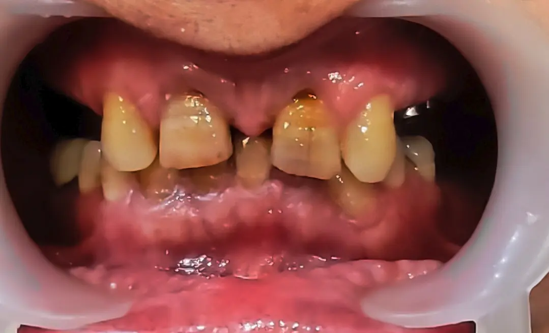
I had a patient whose two upper central incisors had almost no root left and were mobile. There was a large diastema between the two, mostly on the mesial side. The same was seen distally, but less severe. Both lateral incisors were also missing, so the whole span between the two canines was filled by these two centrals and the empty spaces around them.
The treatment plan that had been proposed was to extract the two centrals and place two implants carrying four teeth in this span. I suggested to the patient that before doing anything we should do a mock-up, and I explained what a mock-up is. I also gave the reason: if these two teeth are extracted and four teeth are placed in this span, the final appearance may be so unfamiliar to them that they regret it, while there is no way back any more. On the other hand, although these two centrals have very short roots and an unattractive appearance, they may well remain if we leave them alone. So it makes sense for the patient to see the approximate result before an irreversible decision.
The patient asked whether I could not show on a photo what the result would look like. That question made me think of using AI to build an image mock-up on their own intraoral photo, so they could see the approximate appearance of four teeth in the span between the two canines.
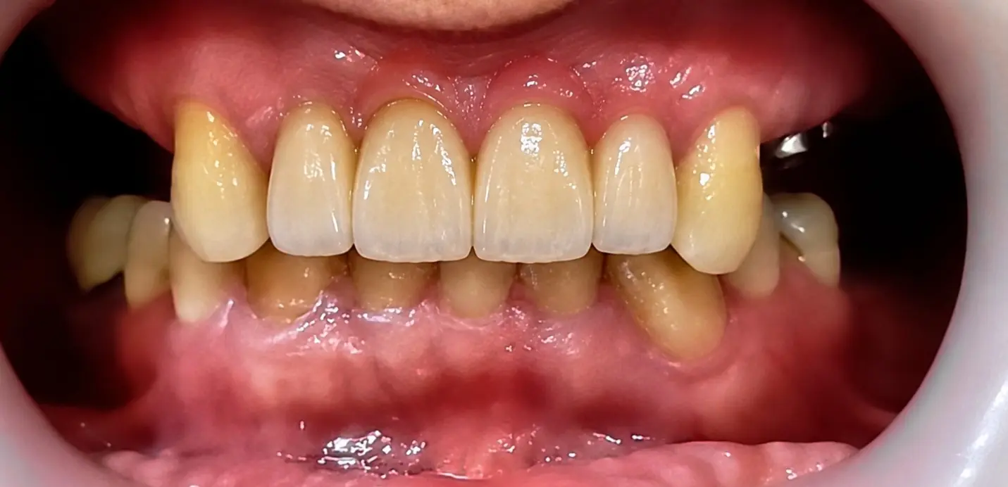
The first image is the patient's current condition and the second is that image mock-up. If in the future the space turns out to be short for four teeth, I can also reduce the mesial of the canines a little.
The content of this page is intended for the educational use of dentists and dental students.