
Jaw Clicking and the Repositioning Splint: From Diagnosis to Treatment
The temporomandibular joint disc lies between the condyle and the articular surface of the temporal bone and helps coordinate joint movement and distribute load. If the position of the disc relative to the condyle changes, the relationship between the two may be disturbed during mouth opening and closing.
In disc displacement with reduction, when the mouth is closed the disc is not in its normal position relative to the condyle; but as the mouth opens, the disc–condyle relationship is re-established. This can be accompanied by a click. On closing, the disc may be displaced again and produce another click. So "with reduction" means that the disc–condyle relationship is re-established during movement, not that the displacement has been resolved permanently.
In disc displacement without reduction, this relationship is not re-established even when the mouth opens. The patient may have limited opening or a feeling that the jaw is locked, although with time and joint adaptation the movement limitation can decrease. So the disappearance of a click is not by itself a sign of improvement, especially if the mouth opens less than before at the same time.
One clinical finding in patients with clicking is elimination of the sound when the mandible is brought forward. The patient is asked to bring the mandible slightly forward and open and close the mouth in that position. If the click is eliminated, this position can serve as the basis for designing an anterior repositioning splint.
But before fabricating a splint, it must be established whether, in addition to the sound, the patient has joint pain or dysfunction. A click without pain or dysfunction does not necessarily need treatment. Also, if the pain is of muscular origin and the joint click is painless, treating the disc displacement will not necessarily resolve the cause of the patient's pain.
The rationale of treatment with a repositioning splint
The anterior repositioning splint guides the mandible into a more forward position on closing. This change can improve the disc–condyle relationship and reduce the load on the painful tissues behind the disc.
In Okeson's account, this temporary position provides an opportunity for the retrodiscal tissues to adapt. After this adaptation, the joint may function without pain, even if the disc is still displaced and the click remains. So the goal of treatment should not be seen only as permanently restoring the disc or eliminating the sound for good.
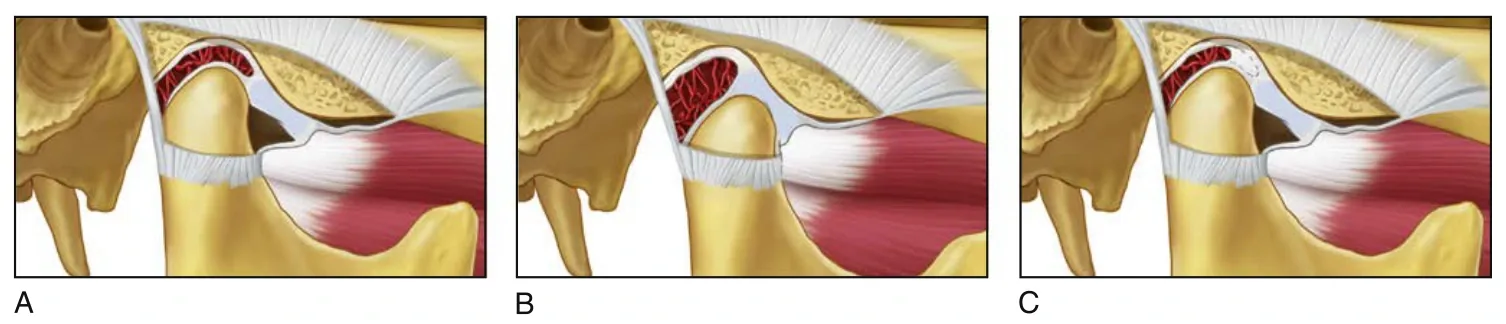
Okeson recommends that if a stabilization splint can control the symptoms, it should be preferred, because it carries a lower risk of occlusal changes. If it is not sufficient, an anterior repositioning splint is considered.
1. Examination and recording the baseline
Pain, joint and muscle tenderness, sounds, a sensation of catching or locking, the range of mandibular movement, and occlusal contacts are examined and recorded. These findings are the basis for comparison at later visits.
Pain on movement or palpation of the joint must be distinguished from muscle pain. It also needs to be established whether the patient's chief complaint is pain, functional limitation, or merely concern about the joint sound.
2. Determining the therapeutic position
The patient is asked to bring the mandible slightly forward and open and close the mouth in that position. According to Okeson, the least protrusion that eliminates the click is the clinical guide for choosing this position. In this position, joint symptoms during opening and closing are also checked.
Elimination of the click is a useful clinical reference for starting treatment, but it does not by itself prove that the disc has returned completely to its normal position.
3. Fabricating and adjusting the splint
In the method described in the book, the splint is made of hard acrylic and covers the entire dental arch. The maxillary arch is usually preferred, because fabricating the guiding ramp that controls the mandibular position is easier on it.
First, an anterior stop is created on the splint. The position in which bringing the mandible forward eliminates the symptoms is marked on this stop with articulating paper. A small groove is then made at the contact site so that the patient can repeat the same position while the contacts are being recorded.
Next, without covering the anterior stop, acrylic is added to the occlusal surface of the splint. The patient closes in the recorded forward position so that the contact sites of the mandibular teeth form in the acrylic. More acrylic is also placed behind the contact sites of the mandibular anterior teeth to create an inclined surface. This surface is the guiding ramp: on closing, the mandibular anterior teeth slide along it and the mandible is guided into the therapeutic position.
After the acrylic has set, the contacts are adjusted so that the teeth have even, simultaneous contacts in this position.
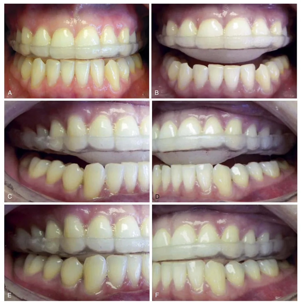
The ramp must be smooth and polished so that the teeth do not catch while moving along it. It is also checked that the splint is stable and retentive, compatible with the soft tissues, and that in the recorded position the symptoms during opening and closing are eliminated.
4. Starting use and follow-up
In the approach of Okeson's 2020 edition, the patient usually starts by wearing the splint during sleep. If daytime use is also needed to control pain, that time is added, and once the pain resolves, use is again limited to the night.
The first follow-up should be early. In the book's clinical example for disc displacement with reduction, the patient was examined one week after the splint was delivered. In the section on stabilization splint follow-up, an initial visit within two to seven days is also mentioned.
At later examinations, pain and tenderness of the joint and muscles, mandibular movement, the condition of the appliance and its contacts are re-examined. Tooth contacts after the splint is removed also matter, because using the appliance can be accompanied by occlusal change.
The interval and continuation of follow-up are set according to the patient's response. If the symptoms do not improve, the fit, the appliance contacts and the way it is being used are checked, and the diagnosis should also be reassessed.
5. When and on what condition to end repositioning
Reducing the use of the splint begins once the pain has resolved. Okeson explains that adaptation of the retrodiscal tissues may take eight to ten weeks or longer. The acuteness and extent of the injury, and the patient's age and general health, affect this time; so the end of treatment is not set simply by reaching a particular date.
In Okeson's older study, after eight weeks of initial treatment, the splint was gradually modified until the original occlusal condition was re-established. This method is called the step-back.
In the book's 2020 edition, the way out of treatment is described mainly through reducing wear time and, if needed, converting the repositioning splint into a stabilization splint. The step-back of the older study and this approach should not be regarded as a single program with fixed time stages.
6. What does modifying or converting the splint mean?
With the help of the anterior ramp, the repositioning splint guides the mandible forward on closing. In contrast, the stabilization splint is adjusted in CR, so that when the condyles are in the musculoskeletally stable position, the teeth have even, simultaneous contacts with the splint.
So converting a repositioning splint into a stabilization splint means that the guiding surface is changed and the contacts on the appliance are readjusted, so that the mandible is no longer guided into the forward position and the contacts are established in CR. In this situation, the repositioning splint is converted into a stabilization splint so that the condyles can return to the musculoskeletally stable position.
According to Okeson, this conversion is one of the ways that allows the condyle to return to the musculoskeletally stable position. After that, the harmony of this position with the tooth contacts is assessed. The phrase "return to the original occlusion" in the older study should also not be equated with reaching CR without examination.
7. If pain returns as use is reduced
According to Okeson, the first and most common explanation is that the tissues have not yet had enough time to adapt. In that case, use of the repositioning splint is reinstated and more time is allowed for adaptation. Once the pain is controlled, reducing use is reconsidered.
If repeated attempts to discontinue the appliance are accompanied by a return of symptoms, orthopedic instability is suspected. In this situation, converting the splint into a stabilization splint and assessing the relationship of the stable joint position to the tooth contacts are part of the decision-making.
Treatment outcome: pain and function alongside sound
In Okeson's 1988 study, after eight weeks, 80% of patients were free of pain and joint sounds. At follow-up averaging two and a half years, 75% had no pain, but joint sounds had returned in 66%. This difference shows that the durability of pain reduction and the durability of click elimination are not the same.
In the Conti study, too, repositioning reduced pain earlier, but at longer follow-up the difference between the groups diminished. In that study, after three to four months of nighttime use, the splint was converted into a stabilization splint at once; so its protocol differed from the step-back of Okeson's study.
During treatment, changes in tooth contacts and posterior open bite should be monitored. Pain reduction, adequate mandibular movement and acceptable function are the main criteria for evaluating the outcome. A painless click that remains does not by itself mean treatment failure or a need to continue repositioning.
∆ References
- Okeson JP. Management of Temporomandibular Disorders and Occlusion. 8th ed. Elsevier; 2020.
- Okeson JP. Long-term treatment of disk-interference disorders of the temporomandibular joint with anterior repositioning occlusal splints. J Prosthet Dent. 1988.
- Conti PCR, et al. Partial time use of anterior repositioning splints in the management of TMJ pain and dysfunction: a one-year controlled study. J Appl Oral Sci. 2005.