Insight 37 — Distal Chipping of the Lateral Veneer: a Hidden Functional Interference in Protrusive Movements
Published:
Last reviewed:
فارسی
Clinical explanation
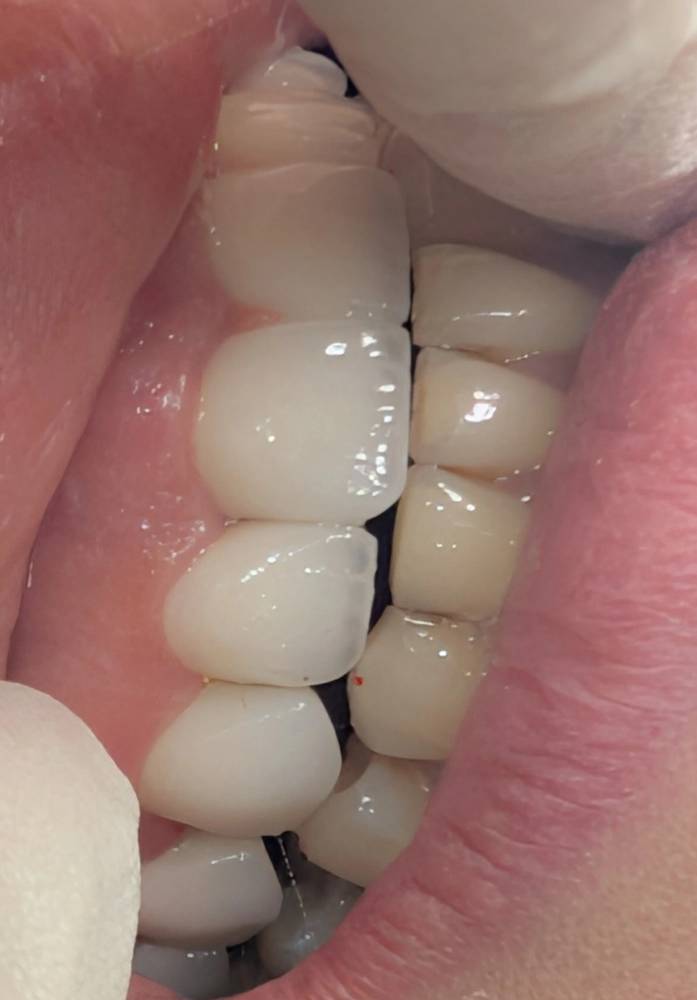
- In delivering anterior veneers, one of the points prone to ceramic fracture is the distal area of tooth 2 (the maxillary lateral); especially when the occlusal adjustments have not been performed precisely and dynamically.
- In this case, the distal area of the maxillary lateral is exposed to direct contact with the mandibular canine. This contact occurs not in maximum intercuspation, but in protrusive movements and the paths of anterior guidance (Anterior Guidance).
- The point is that these kinds of interferences are often not seen in a static examination and reveal themselves only in movement.
-
Two key factors play a role in creating this problem:
- 1. Incorrect setting of the anterior Guidance — if the movement path of the anterior teeth has not been correctly designed and adjusted, the lower canine can, while moving forward, strike the distal area of the maxillary lateral.
- 2. Incorrect design of the distal lateral contour — a prominence or over-contour in this area can act as a premature contact point and transmit force directly to the edge of the ceramic.
- The consequence of this interference is force being applied to the thin edge of the ceramic, which in the long term — or even the short term — leads to chipping (Chipping).
- Key point: evaluating occlusion in anterior veneers is not limited to static contacts. Each unit must also be evaluated in the laterotrusive and protrusive movement path. The distal of the maxillary lateral is one of the points that, if not seen, reveals itself with failure.
Ceramic fracture at the distal of the lateral; the consequence of overlooking the movement path of the lower canine
The content of this page is intended for the educational use of dentists and dental students.