
Permanent Printed Crowns: How Much Can We Trust Them?
In full-arch implant rehabilitation, one important challenge is accurately recording the positions of the implants relative to one another. This matters because even a small recording error in a screw-retained prosthesis can create strain when the screws are tightened and compromise passive fit.
In a conventional digital workflow, scan bodies are attached to the implants or multi-unit abutments, and their positions are recorded with an intraoral scan.
However, full-arch scanning in a completely edentulous patient is not always reliable. The lack of stable landmarks, the distance between implants, and possible errors while the scanner stitches sequential images together can make the recorded positions differ from the implants’ actual positions.
One approach proposed to address this problem is the Reverse Scan Body.
Where did the idea come from?
To understand the rationale, it helps to first look at how a full-arch provisional prosthesis is made.
Suppose a provisional prosthesis has been designed and made for a patient who is going to receive full-arch treatment. It may have been based on an initial scan, a digital design, and the planned implant positions.
But the implant positions after surgery are not necessarily identical to the planned positions. Differences may remain even when guided surgery is used.
In one common method for making a provisional prosthesis, temporary cylinders are first secured to the multi-unit abutments. The provisional, with enough space for those cylinders to pass through, is then positioned in the mouth.
Once the prosthesis is in the right position, the cylinders are connected to it with resin. This step is commonly called an Intraoral Pickup.
After the resin polymerizes, the prosthesis is removed and its fit is checked. If needed, the connections are corrected or remade so that passive fit can be confirmed.
A provisional prosthesis that has gone through pickup and fit verification therefore contains information that may not have been present in the initial design or scan: the actual positions of the implant connections relative to one another.
This is only one way to make a provisional. Other methods may start with an accurate impression or model. For a Reverse Scan Body workflow, what matters is having a prosthesis with verified fit.
What does a Reverse Scan Body do?
When a provisional prosthesis has passive fit, it can be used to transfer implant positions into the digital environment.
In the Reverse Scan Body method, the provisional is removed from the mouth, and special scan bodies are attached to the connections on its intaglio surface.
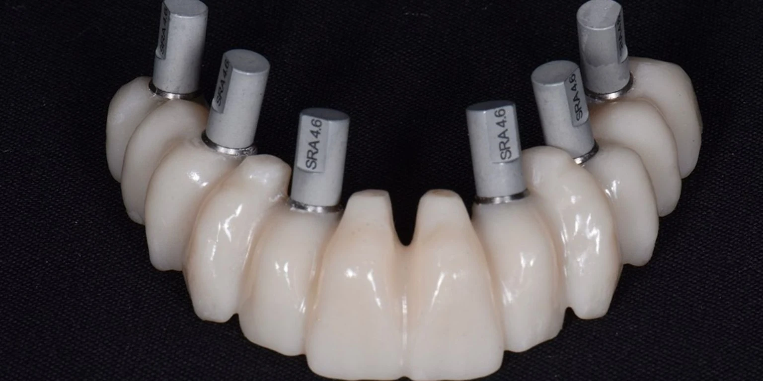
The prosthesis and scan bodies are then scanned outside the mouth.
Using the known geometry of the scan bodies and their digital library, the software can reconstruct the three-dimensional positions of the implant connections.
Put simply, instead of recording implant positions directly inside the mouth, we extract them from a prosthesis that has already been fitted to those same implants and whose fit has been verified.
This is the key difference between a Reverse Scan Body and a conventional scan body.
Why is it called “Reverse”?
In a conventional method, the scan body is placed on an implant or abutment in the mouth and scanned to identify the prosthetic connection’s position.
In the Reverse method, the scan body is attached to the prosthesis itself, and the implant positions are reconstructed from the prosthesis side.
The direction of information transfer is reversed.
The concept is not entirely new. In analog workflows, a well-fitting provisional prosthesis could also be used to transfer implant positions and make a model. This method is known as Back-Pouring.
In that method, the corresponding analogs were connected to the abutments and fixed inside the prosthesis. Stone was then poured to make a cast that reproduced the implant connections’ positions.
Reverse Scan Body can be considered the digital equivalent of this concept.
What information do we obtain?
One advantage is that implant positions can be recorded together with the shape of the provisional prosthesis in a single digital dataset.
If tooth positions, occlusion, and the overall form of the provisional have already been evaluated and approved in the mouth, these data can help guide the design of the definitive prosthesis.
When needed, an intraoral scan of the prosthesis, a scan of the opposing arch, the interarch relation, and a soft-tissue scan can also be taken. These data are aligned with the extraoral scan of the prosthesis in the software.
So the Reverse Scan Body is not only a tool for recording implant positions; it can also help transfer information from a verified provisional prosthesis into the design stage for the definitive prosthesis.
Is this method always more accurate?
Not necessarily.
A Reverse Scan Body does not correct errors in the provisional prosthesis by itself. If an error occurred during cylinder pickup, if the resin connection distorted, or if the prosthesis did not have adequate passive fit, the same error may be carried into the digital file.
The accuracy of the extraoral scan, the scanner type, and the characteristics of the prosthesis can also affect the result.
That is why verifying the fit of the reference prosthesis is essential.
This should be considered an alternative route for digitally transferring implant positions, not a method that is necessarily more accurate than intraoral scanning or photogrammetry in every situation.
Summary
The Reverse Scan Body concept is based on using information from a prosthesis whose implant connections have already been recorded in the mouth and whose fit has been verified.
Instead of recording implant positions again from inside the mouth, the provisional prosthesis is used as a reference, and its information is transferred into the digital environment through an extraoral scan.
A Reverse Scan Body does not create an accurate position; it transfers into the digital environment a position that has already been recorded and verified.