
When You See a Crack Under Caries or an Old Restoration, How Far Should You Go?
Suppose you are removing an old restoration or caries from a vital tooth and see a crack line on the floor or wall of the cavity. Should you keep cutting until the crack line disappears completely? Or can you leave part of it and seal over it?
On the one hand, we do not want to leave a path open for bacteria to reach the pulp; on the other, we do not want to remove sound dentin for no reason or expose the pulp by cutting further.
Why do we remove the crack?
A crack that communicates with the oral environment gradually fills with bacterial biofilm. Bacteria can also penetrate toward the pulp through the dentinal tubules [3]. For this reason, in a tooth with reversible pulpitis, it is recommended that the crack line be removed carefully and the cavity finally sealed with a bonded restoration [2].
In a vital tooth, how far should we go?
In our search, we did not find a reliable clinical study that sets a specific depth at which cutting should stop.
A small clinical study in 2024 examined exactly this question in vital teeth. In that study, the crack was cut back only to 2 mm into dentin and no further. In some teeth, the crack line was still visible on the cavity floor. These teeth were also restored with composite, and after one year their outcome did not differ significantly from teeth at the same 2 mm depth in which no crack line was visible on the cavity floor [4].
But this study has two limitations. The number of these teeth was very small, and treatment failure was related to the depth of the crack [4]. So 2 mm cannot be called a safe limit for stopping.
On the other hand, a laboratory study in 2022 showed that using a bur to remove a crack on the cavity floor can create new microcracks or extend the existing crack [5]. The cracks examined in that study were artificial [5], but the findings show that chasing a crack to its very last point is not without consequences.
The boundary proposed by the biomimetic approach
In the biomimetic approach, crack removal follows the same principles used for removing deep caries [9].
In this method, the dentin crack is removed completely within the 2 mm band adjacent to the DEJ, the peripheral seal zone, so that the restoration margins rest on sound tissue and proper bonding is possible [9].
In the central part of the cavity, however, removal stops at a depth of 5 mm from the occlusal surface and, in the proximal area, 3 mm from the axial wall [9].
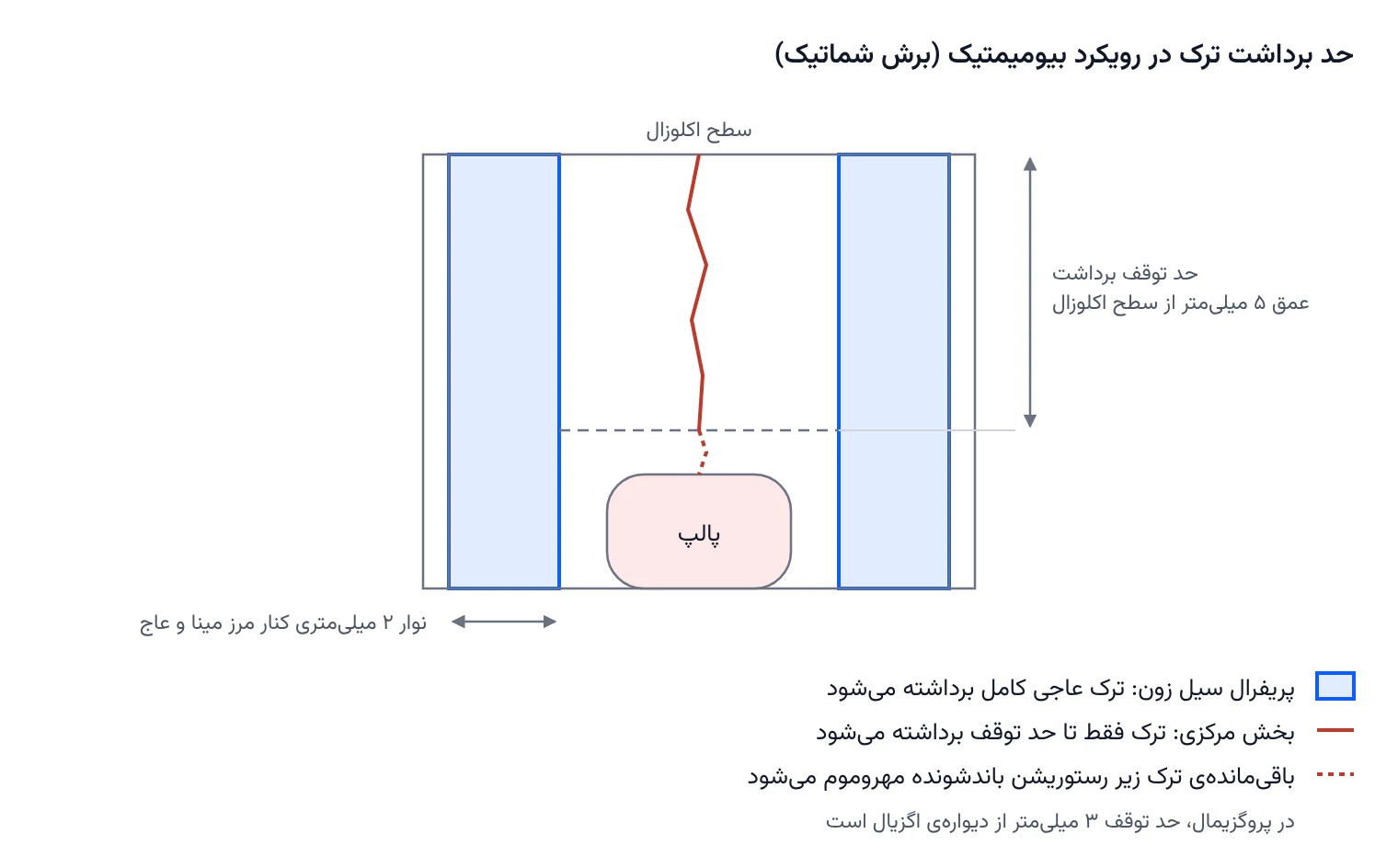
These numbers were set to avoid pulp exposure in a vital tooth [8]. They are based on tooth anatomy, not on clinical studies of treatment outcomes in cracked teeth [8]. A 2024 critical review also stressed that biomimetic protocols need closer scrutiny and more rigorous research [10].
The biomimetic approach and the limited removal described in the previous section agree on one point: in the central part of the cavity we should not keep cutting toward the pulp to chase the crack. What the biomimetic approach adds is the cavity periphery: this zone must be completely free of cracks so that the restoration bonds to sound tissue.
After the crack is removed, pulp status determines the decision
A crack on its own does not mean that root canal treatment is needed.
In 2009, Abbott and colleagues treated vital cracked teeth with reversible pulpitis using a conservative approach. After the crack, caries and the previous restoration were removed, a liner and an interim restoration were placed and pulp status was followed. About 80 percent of these teeth did not need root canal treatment [1].
Root canal treatment was done from the outset in cases where the pulp was exposed, the crack reached the pulp, or the remaining tooth structure was such that its rebuilding required a post [1].
If the patient still has pain on chewing after the cavity is closed
In this situation we should think about covering the cusps. One recommendation is that, if pain on chewing persists, the tooth be protected with a provisional crown and the need for root canal treatment then be re-evaluated [2].
Covering the cusps does not necessarily require a full crown, though. An onlay, or even composite cusp coverage, can achieve the same goal while removing less tooth structure.
In a large study with three-year follow-up, treatment failure was associated with intracoronal restorations, not with restorations that covered the cusps fully or partially [11]. Put simply, in our view the key point is to cover the cusps so that the two sides of the crack do not move independently of each other under chewing forces.
If the pain resolves with cusp coverage, restorative treatment can continue. But if the pain persists or signs of pulpitis appear, pulp status should be reassessed and root canal treatment done if needed.
Cuspal coverage: why does it matter so much?
The final restoration of a symptomatic or root-canal-treated cracked tooth should be a full crown. A 2024 meta-analysis examined treatment outcomes separately for vital teeth and root-canal-treated teeth [6].
In vital teeth, direct restorations without cuspal coverage were compared with full crowns. The likelihood of pulpal complications in the group without cuspal coverage was about 3 times higher [6]. A higher likelihood of extraction was also reported in this group, although its numerical estimate was not reliable enough [6].
In root-canal-treated teeth, the difference was larger. The likelihood of extraction for root-canal-treated cracked teeth without full crowns was reported to be about 11 times higher [6].
If the tooth needs root canal treatment
In cases of irreversible pulpitis or pulp necrosis, root canal treatment is done first, and the tooth is then protected with a provisional crown so that, once symptoms resolve, a definitive crown is made [2].
After the access cavity is opened, we can assess the extent of the crack better. If the crack runs from one side of the pulp chamber floor to the other or extends into a canal, the prognosis of the tooth will be poor [7].
In a survey of American endodontists, most specialists in this situation, especially in the presence of a 6 mm periodontal pocket, chose to extract the tooth [7]. This study, however, shows the specialists' decisions, not the actual outcome of treating these teeth.
A sign that is worrying in both cases
If the crack has extended below the gingiva and is associated with a deep periodontal pocket, the prognosis of the tooth is unfavorable, whether the pulp is vital or the tooth has already had root canal treatment. It is best to discuss this with the patient before continuing treatment [2].
The decision at a glance
| Situation | Action | Source |
|---|---|---|
| Vital tooth with a normal pulp or reversible pulpitis | Conservative crack removal, a bonded restoration and pulp follow-up | [1], [2] |
| Pain on chewing after the cavity is closed | Cusp coverage (onlay or crown) and pulp re-evaluation | [2], [11] |
| Exposed pulp or a crack reaching the pulp | Root canal treatment | [1] |
| Irreversible pulpitis or pulp necrosis | Root canal treatment, a provisional crown, then a definitive crown | [2], [6] |
| Crack across the pulp chamber floor or into a canal | Poor prognosis; most specialists chose extraction | [7] |
| Subgingival crack with a deep periodontal pocket | Unfavorable prognosis; discuss with the patient before continuing | [2] |
Summary
In a vital tooth, the cavity periphery should be cleared of cracks so that the restoration margins rest on sound tissue and good bonding is achieved. In the central part of the cavity, however, we should not go toward the pulp to remove the crack line completely. We can stop removal at a conservative limit and seal the remaining part under a bonded restoration.
After that, pulp status determines whether root canal treatment is needed. Finally, by covering the cusps, we protect the tooth against chewing forces.
∆ References
- Abbott et al., Aust Dent J, 2009 (abstract)
- Kim, Restor Dent Endod, 2016
- Chen et al., J Dent, 2023 (abstract)
- Shrestha, IJISRT, 2024 (abstract)
- Hovander et al., JADA Found Sci, 2022 (abstract)
- Zhang et al., J Dent, 2024 (abstract)
- Abulhamael et al., J Contemp Dent Pract, 2019 (abstract)
- Alleman and Magne, Quintessence Int, 2012 (abstract)
- DentCast's Alleman biomimetic course: crack dissection and crack removal end points (CrRE)
- Reis et al., J Appl Oral Sci, 2024 (abstract)
- Ferracane et al., Clin Oral Investig, 2021 (abstract)